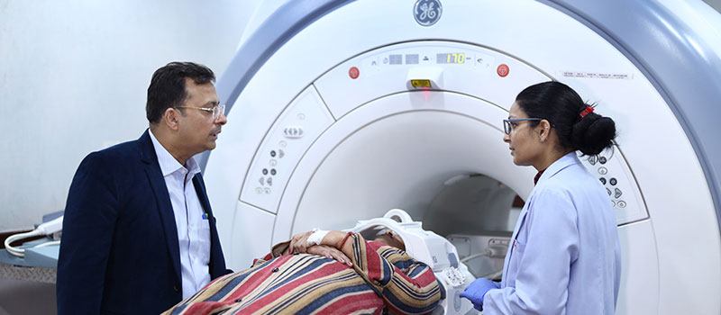
MRI
Magnetic Resonance Imaging (MRI) is one of contemporary medicine's most effective imaging techniques. Unlike many imaging technologies, the MRI creates images using magnets rather than radiation. In addition to being painless and noninvasive, MRIs are currently one of the most effective and efficient imaging examinations accessible.
A patient may be told to get an MRI for several reasons, such as:
Types of MRIs
Several types of MRIs are offered by Dr. Sanjay Gupta at Health Care Imaging Centre in Shivaji Marg in Meerut, Uttar Pradesh, including:
MRI Shoulder
Shoulder MRI involves using a strong magnetic field, radio waves, and a computer to create high-resolution images of the shoulder's skeletal structure, soft tissues, and blood arteries. Most often, it is used in the process of injury evaluation.
MRI Elbow
In an elbow MRI by Dr. Sanjay Gupta at Health Care Imaging Centre, Meerut patients are positioned supine or prone with the arm. The imaging starts around 10 centimetres above the elbow and continues to the bicipital tuberosity.
MRI Wrist
A systematic review of the MRI of the wrist is very important because the anatomy of the wrist is very complicated, with many small structures, diseases, and injury patterns that can be treated in many different ways.
MRI Hand
During the MRI at Health Care Imaging Centre, you will be put on a table in the middle of a large scanner that looks like a tube but has holes at both ends. The hand or finger that is hurt will then be put in an MRI coil.
MRI HIP
An MRI may detect cartilage and labrum tears and fray. Occasionally, it is vital to distinguish between pain emanating from the hip joint and discomfort emanating from the lower abdomen. To do this, a steroid analgesic may be injected into the hip.
MRI Knee
When Dr. Sanjay Gupta does an MRI at Health Care Imaging Centre, Meerut, on the knee, a strong magnetic field, radio waves, and a computer are used to make detailed pictures of the structures inside the knee joint. Usually, it is used to help find the cause of pain, weakness, swelling, or bleeding in and around the joint or to figure out how bad it is. Knee MRI doesn't use ionizing radiation and can help you figure out if you need surgery.
MRI Ankle
MRIs can show ligament injuries and have been used to tell ligament tears apart from other causes of ankle pain, like a broken bone, an injury to the cartilage, or a torn tendon.
MRI Foot
Using strong magnets and radio waves, an MRI foot scan may provide clear pictures of the foot's skeletal structure, including the muscles, tendons, and ligaments, and it causes no discomfort to the patient.


MRI Preparations
You don't have to do much to prepare for an MRI scan. You won't get any radiation from the test, so it's safe and won't hurt you. Before getting an MRI, you should talk to Dr. Sanjay Gupta at Health Care Imaging Centre, Meerut, about the following:
Tell Dr. Sanjay Gupta if you have metal implants because they could affect the test results. MRIs don't use radiation; instead, they use magnetic fields. This means that they can be used with most types of metal. Among other things:

Please keep in mind that everyone will have to change before their MRI.
You can choose where you want to get your MRI done. Health Care Imaging Centre has some of the most advanced technology. If you want to know more, you can call us at 08923000078.
MRI FAQs
Even though MRIs and CT scans can make similar images, the MRI is usually thought to be the more powerful of the two. But MRIs take longer and cost more than other tests. Also, MRIs don't use X-rays. Instead, they use magnetic fields so patients don't get any radiation.
Even though MRIs are thought to be more powerful, CTs and MRIs can be better for taking pictures of different body parts. CT scanning is usually the best way to look at the lungs, while MRIs are usually better for knee MRIs, foot MRIs, and shoulder MRIs, to name a few. MRIs are also often used to clear up CT scan results that aren't clear enough.
The length of an MRI can range from 15 to 90 minutes. About 30 minutes is the average time.
You will not get your test results immediately after completion. Radiologists need sufficient time to analyse the pictures and compare them to your medical records and any prior images that may be on file. They normally report their findings to the referring physician within 48 hours, who will contact you to discuss the results.
